Very rarely do people still buy horses without a valid veterinary check-up. Even at lower levels, it can be important to have a veterinarian examine the horse you are interested in buying. Oftentimes buyers will want a set of X-rays of the horse's limbs and/or back and neck to assess the risks of future injuries. But how do X-rays work? And what do they show?
What are x-radiation waves?
X-radiation waves are electromagnetic waves. Radio waves, infrared waves which produce heat and light waves which produce visible light also belong in to the group of electromagnetic waves. Compared to these however, x-rays have a much shorter wavelength and are produced inside an x-ray tube, made of either glass, metal or ceramics, which contains a vacuum. To produce x-rays, an electrical current is sent through a cathode, which is a negatively charged metal filament, to heat it to a very high temperature. This increased heat releases small negatively charged particles, called electrons. These electrons travel through the vacuum at high speed towards an anode, a positively charged part of metal. The collision of the electrons with the anode produces a huge amount of energy: mostly as heat, but some as x-rays. These x-rays leave the x-ray tube as a beam through a series of lead shutters.
How are x-rays captured?
Originally, images were captured on an x-ray film combined with and intensifying screen which converts x-rays into light signals and makes them visible to the naked eye. However, in this digital day and age this has been replaced by digital capture devices, usually in protective containers called cassettes. Parts of the body where the tissue is very dense absorb a lot of x-rays and therefore appear pale or white on the x-ray film, whereas more x-rays pass through the parts of the body where tissue is less dense and expose the film. This means that when x-rays pass through air, the image will appear black and when they pass through soft tissue or other fluids, it will show up in varying shades of grey.
What is x-ray imaging used for?
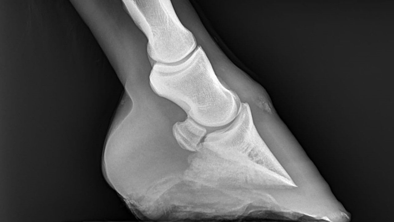
The capture of x-rays or a radiographic examination of horses is mainly used to examine the structure of bones. However horses are large animals, which means that the radiation required to capture these images is much higher compared to humans. Muscle tissue absorbs x-rays, therefore some parts of the horse's body are particularly hard to visualise radiographically: thoracic and lumbar spine, the scapula, the pelvis and the femur. Other bones and joints of the horse's body are more easily captured, such as the skull, the cervical spine or neck, the shoulder joint, elbow joint, carpal joint, stifle joint and tibia, hock joint, the cannon bone and splint bones, the digit and the hoof.
How are x-rays interpreted?
The most important part of an x-ray examination is the interpretation of the images. The person who interprets the radiographs must be able to correctly identify all the anatomical structures that are visible on the image, therefore they need to have an extensive knowledge of equine anatomy, which is why it is usually done by a veterinarian. The quality of the radiograph is also very important as well as being able to identify things that are not part of the horse's anatomy like dirt or trapped air.
What are the most common structural changes found in x-ray images?
Structural changes are most commonly known as pathologies, most typically caused by trauma or disease. There is a large number of pathologies that can appear on a radiograph which is why the interpretation is best left to a specialist veterinarian or radiologist. However, some structural changes show up more often commonly than others. For example, uneven or proliferated margins of a bone, which appear most often around the joints. Fractures or fissures are pictured on an x-ray as loss of continuity of that bone. Uneven, narrowed or enlarged joint spaces are typical findings, as well as fragments of bones at the joint margins, popularly known as OCD chips. Cysts, which often appear directly adjacent to a joint, show up as circular areas of less dense bone, meaning they appear darker on the x-ray. Other common changes are mineralisation of soft tissues, such as tendons and ligaments, as well as foreign objects or air pockets trapped in soft tissue or fluid found in cavities which should be air-filled (sinuses).
Although these findings are relatively common, they are not easy to recognise. It is important to not 'misinterpret' a normal anatomical feature as something abnormal. When there is a doubt, it may be smart to get a second opinion by a specialised veterinarian radiologist. Even then it is incredibly hard to predict how a particular horse will react to a specific change in a radiograph. Veterinarians, unfortunately, do not have a crystal ball and as every horse is unique, they cannot 100% predict the outcome.